LIPIDS AND FATTY ACIDS

Lipids: Fats, Oils, Waxes, etc.

What Are Lipids?

All Lipids are hydrophobic: that’s the one property they have in common. This group of molecules includes fats and oils, waxes, phospholipids, steroids (like cholesterol), and some other related compounds.

Structure of Fatty Acids

One Fatty Acid
One Fatty Acid
The “tail” of a fatty acid is a long hydrocarbon chain, making it hydrophobic. The “head” of the molecule is a carboxyl group which is hydrophilic. Fatty acids are the main component of soap, where their tails are soluble in oily dirt and their heads are soluble in water to emulsify and wash away the oily dirt. However, when the head end is attached to glycerol to form a fat, that whole molecule is hydrophobic.

Fatty Acids
Fatty Acids
The terms saturated, mono-unsaturated, and poly-unsaturated refer to the number of hydrogens attached to the hydrocarbon tails of the fatty acids as compared to the number of double bonds between carbon atoms in the tail. Fats, which are mostly from animal sources, have all single bonds between the carbons in their fatty acid tails, thus all the carbons are also bonded to the maximum number of hydrogens possible. Since the fatty acids in these triglycerides contain the maximum possible amouunt of hydrogens, these would be calledsaturated fats. The hydrocarbon chains in these fatty acids are, thus, fairly straight and can pack closely together, making these fats solid at room temperature. Oils, mostly from plant sources, have some double bonds between some of the carbons in the hydrocarbon tail, causing bends or “kinks” in the shape of the molecules. Because some of the carbons share double bonds, they’re not bonded to as many hydrogens as they could if they weren’t double bonded to each other. Therefore these oils are called unsaturated fats. Because of the kinks in the hydrocarbon tails, unsaturated fats can’t pack as closely together, making them liquid at room temperature. Many people have heard that the unsaturated fats are “healthier” than the saturated ones. Hydrogenated vegetable oil (as in shortening and commercial peanut butters where a solid consistency is sought) started out as “good” unsaturated oil. However, this commercial product has had all the double bonds artificially broken and hydrogens artificially added (in a chemistry lab-type setting) to turn it into saturated fat that bears no resemblance to the original oil from which it came (so it will be solid at room temperature).

Cis and Trans Bonds
Cis and Trans Bonds
In unsaturated fatty acids, there are two ways the pieces of the hydrocarbon tail can be arranged around a C=C double bond. In cis bonds, the two pieces of the carbon chain on either side of the double bond are either both “up” or both “down,” such that both are on the same side of the molecule. In trans bonds, the two pieces of the molecule are on opposite sides of the double bond, that is, one “up” and one “down” across from each other. Naturally-occurring unsaturated vegetable oils have almost all cis bonds, but using oil for frying causes some of the cis bonds to convert to trans bonds. If oil is used only once like when you fry an egg, only a few of the bonds do this so it’s not too bad. However, if oil is constantly reused, like in fast food French fry machines, more and more of the cis bonds are changed to trans until significant numbers of fatty acids with trans bonds build up. The reason this is of concern is that fatty acids with trans bonds are carcinogenic, or cancer-causing. The levels of trans fatty acids in highly-processed, lipid-containing products such as margarine are quite high, and the government now requires that the amounts of trans fatty acids in such products be listed on the labels.

Omega-3 and Omega-6
Omega-3 and Omega-6 Fatty Acids
Another set of fat-related terms that are “in the news” a lot, lately are “omega-3” and “omega-6” fatty acids. These terms both refer to fatty acids that have at least one unsaturated bond in their chains, and these terms describe where that bond is located. Starting from the end of the carbon chain (that does not contain the carboxyl group and is not bonded on to the glycerol), the carbon atoms before the first double bond are counted. If there are three carbons, it is an omega-3 fatty acid, and if there are are six carbons, it is an omega-6 fatty acid. We need a balanced amount of both in our diets, but the omega 3 fatty acids are not as common, thus harder to obtain, and so many people’s intake of these two types of fatty acids is way out of balance, including way too many omega-6 fats as compared to the amount of omega-3 fats in their diets. Flax seed and chia seed contain significant amounts of omega-3 fatty acids. Certain types of marine algae manufacture lots of omega-3 fatty acids, and those are incorporated into the tissues of fish which eat those algae. Because of that, many people take fish-oil capsules to increase the amount of omega-3 fats in their diets, but especially for vegetarians, consuming algae-derived omega-3 fats is another option.

Structure of Fats and Oils

Glycerol
Glycerol
Triglyceride
Triglyceride
Fats and oils are made from two kinds of molecules: glycerol (a type of alcohol with a hydroxyl group on each of its three carbons) and threefatty acids joined by dehydration synthesis. Since there are three fatty acids attached, these are known as triglycerides. “Bread” and pastries from a “bread factory” often contain mono- and diglycerides as “dough conditioners.” Can you figure out what these molecules would look like? The main distinction between fats and oils is whether they’re solid or liquid at room temperature, and this, as we’ll soon see, is based on differences in the structures of the fatty acids they contain.

The fatty acids that make up the fats and oils in our diets and our bodies may also be lumped into groups based upon the number of carbon atoms in their chains. Interestingly, most of the fatty acids in living organisms have an even number of carbons (2, 4, 6,…). Fatty acids with less than 6 carbons in their chains may collectively be called short-chain fatty acids, but usually these are just called carboxylic acids and are not referred to as “fatty” acids. Those with 6 to 12 carbons are medium-chain fatty acids, those with 14 to 22 carbons are long-chain fatty acids, and those with over 22 carbons are thevery-long-chain fatty acids. Since, as mentioned above, fats and oils contain three fatty acids and are called triglycerides, those which contain primarily medium-chain fatty acids are referred to as medium-chain triglycerides (MCTs), while those which contain primarily long-chain fatty acids are referred to as long-chain triglycerides (LCTs). Most of the fats and oils in our diets and that we’re used to hearing about in the news are LCTs.

Chain Length # of Carbons Name Formula Notes
The first 11 saturated fatty acids are:
short-chain C2 acetic acid CH3COOH aceto = vinegar
C4 butryic acid CH3(CH2)2COOH butyr = butter
medium-chain C6 caproic acid CH3(CH2)4COOH capri = goat
C8 caprylic acid CH3(CH2)6COOH
C10 capric acid CH3(CH2)8COOH
C12 lauric acid CH3(CH2)10COOH lauri = laurel
long-chain C14 myristic acid CH3(CH2)12COOH myrist = anoint, ointment
C16 palmitic CH3(CH2)14COOH
C18 stearic acid CH3(CH2)16COOH stear = fat, suet, tallow
C20 arachidic or eicosanoic acid CH3(CH2)18COOH arachis = a leguminous plant, present in peanut oil
C22 behenic or docosanoic acid CH3(CH2)20COOH present peanut & canola oils
A few of the more important unsaturated fatty acids include:
EFA = essential fatty acid, PUFA = polyunsaturated fatty acid, MUFA = monounsaturated fatty acid.
(CH2)7, for example, means repeat CH2 seven times in a row, so CH2CH2CH2CH2CH2CH2CH2.
To determine the omega number, count the number of carbons in from the left end of each of
the following molecules until the first double bond. Do not start counting from the carboxyl group.
C18 oleic acid CH3(CH2)7CH=CH(CH2)7COOH omega-9, MUFA, present in olive & sesame oils, oleo = olive, olive oil
C18 linoleic acid CH3(CH2)4CH=CHCH2CH=CH(CH2)7COOH EFA, omega-6, PUFA
C18 linolenic or alpha linolenic acid (ALA) CH3CH2CH=CHCH2CH=CHCH2CH=CH(CH2)7COOH EFA, omega-3, PUFA, present in flaxseed oil
C20 arachidonic acid (ARA) CH3(CH2)4(CH=CHCH2)4(CH2)2COOH EFA, omega-6, PUFA
C20 eicosapentaenoic or timnodonic acid (EPA) CH3(CH2CH=CH)5(CH2)3COOH omega-3, PUFA, present in algae/fish oil
C22 docosahexaenoic or cervonic acid (DHA) CH3(CH2CH=CH)6(CH2)2COOH omega-3, PUFA, present in algae/fish oil

Notice that, while these unsaturated fatty acids contain the same number of carbons as some of the saturated fatty acids, they are chemically different and have different names.

The medium-chain triglycerides (MCTs) play some very interesting roles in our bodies. MCTs are handled quite differently by our bodies. Our digestive tract doesn’t need to digest them or even use bile to emulsify them, but they are directly absorbed into our blood and sent to the liver where some of them are converted into ketones. Both the MCTs themselves and the ketones formed from them are directly used by our brains and our muscles as alternate fuel/energy sources in place of glucose. In general, MCTs are not stored as body fat, so unless a person would consume way more than necessary, they do not contribute to weight gain. However, consumption of “too much” is unlikely because eating more at once than one’s body is used to tends to cause diarrhea. Even though they are saturated fats, they are “heart-healthy,” and the heart actually beats more effeciently using them as fuel in place of glucose.

Think what life must have been like back in “cave-man days” — people back then weren’t assured of three sumptous, regularly-scheduled meals a day, but rather, if they hadn’t killed a buffalo or a gazelle in several days, they might have nothing to eat for a couple days. If glucose (sugar) was the only fuel their brains and muscles could use to keep going, they would have, long-ago, starved to death, and we wouldn’t be here today. Rather, being able to use MCTs as an energy source enabled them to keep going and survive. Even today, mothers’ milk is very high in MCTs (10 to 17% of its fat), newborns’ brains use MCTs and their metabolic derivatives for as much as 25% of their energy requirements, and now, every infant formula on the market these days contains MCTs.

In terms of our brains’ abilities to use glucose as a fuel, it turns out that the insulin produced by our pancreas that helps transport sugar into all our other body cells (so it can be used as an energy source) cannot cross the blood-brain barrier, but rather, our brains make their own insulin which is used to help transport glucose into the neurons (nerve cells, brain cells) so they can use it for fuel. In terms of pancreatic insulin, you’ve probably heard of type I and type II diabetes, in which either the person’s pancreas isn’t making enough insulin or else the cells that need to take the sugar out of the blood have “broken” insulin receptors, so even if the person’s pancreas is making enough insulin, that “message” to take sugar out of the blood never gets received by the cells. While we tend to think about that in terms of all the sugar that’s staying in the blood, think for a minute what that means in terms of the cells that aren’t getting the sugar inside when they need it.

Now, instead of the rest of the body, think of all this in terms of the brain cells. Don’t think in terms of blood or in terms of fluid within the brain, but think in terms of the actual brain cells, the neurons. What if a person’s neurons were insulin resistant? That would mean that sugar couldn’t get into the neurons to be used as energy to keep the neurons functioning. The neurons would end up, essentially, starving to death. Does that sound far-fetched or impossible? It turns out, based on current research, that can and does happen. Alzheimer’s is now being referred to as type III diabetes. It has been observed that many people who have been diagnosed with Alzheimer’s or other similar neurological diseases were “sugar junkies” for years before their diagnosis. While these people weren’t showing Alzheimer’s signs and symptoms, yet, their neurons were starving, even then, and were sending out messages “begging” for more sugar. Unfortunately, because those neurons were unable to get all that sugar inside themselves, it didn’t do any good, and they began to die off. When enough neurons had died, and thus, the person’s brain had “shrunk” enough for the deficit to be noticeable, then the person was diagnosed with Alzheimer’s or one of the other, closely-related neurological diseases.

MCTs are also being used to treat some types of cancer. Some very aggressive, rapidly-growing cancers can only use glucose as an energy source. Thus it has been found that, if people with those types of cancer are put on a special diet that is very low in sugar, but has adequate MCTs, the person’s body can use the MCTs for fuel, while the cancer starves.

We need fats in our bodies and in our diet. Animals in general use fat for energy storage because fat stores 9 KCal/g of energy. Plants, which don’t move around, can afford to store food for energy in a less compact but more easily accessible form, so they use starch (a carbohydrate, NOT A LIPID) for energy storage. Carbohydrates and proteins store only 4 KCal/g of energy, so fat stores over twice as much energy/gram as fat. By the way, this is also related to the idea behind some of the high-carbohydrate weight loss diets. The human body burns carbohydrates and fats for fuel in a given proportion to each other. The theory behind these diets is that if they supply carbohydrates but not fats, then it is hoped that the fat needed to balance with the sugar will be taken from the dieter’s body stores. Fat is also is used in our bodies to a) cushion vital organs like the kidneys and b) serve as insulation, especially just beneath the skin.

Phospholipids

Lecithin
Lecithin
Phospholipids are made from glycerol, two fatty acids, and (in place of the third fatty acid) a phosphate group with some other molecule attached to its other end. The hydrocarbon tails of the fatty acids are still hydrophobic, but the phosphate group end of the molecule is hydrophilic because of the oxygens with all of their pairs of unshared electrons. This means that phospholipids are soluble in both water and oil.

Mayo RecipeAn emulsifying agent is a substance which is soluble in both oil and water, thus enabling the two to mix. A “famous” phospholipid is lecithin which is found in egg yolk and soybeans. Egg yolk is mostly water but has a lot of lipids, especially cholesterol, which are needed by the developing chick. Lecithin is used to emulsify the lipids and hold them in the water as an emulsion. Lecithin is the basis of the classic emulsion known as mayonnaise. For more information on mayonnaise, see the Biol 1081L Mayonnaise Web page.

Phospholipid Bilayer
Our cell membranes are made mostly of phospholipids arranged in a double layer with the tails from both layers “inside” (facing toward each other) and the heads facing “out” (toward the watery environment) on both surfaces.

Steroids

Cholesterol
Cholesterol
The general structure of cholesterol consists of two six-membered rings side-by-side and sharing one side in common, a third six-membered ring off the top corner of the right ring, and a five-membered ring attached to the right side of that. The central core of this molecule, consisting of four fused rings, is shared by allsteroids, including estrogen (estradiol), progesterone, corticosteroids such as cortisol (cortisone), aldosterone, testosterone, and Vitamin D. In the various types of steroids, various other groups/molecules are attached around the edges. Know how to draw the four rings that make up the central structure.

Cholesterol is not a “bad guy!” Our bodies make about 2 g of cholesterol per day, and that makes up about 85% of blood cholesterol, while only about 15% comes from dietary sources. Cholesterol is the precursor to our sex hormones and Vitamin D. Vitamin D is formed by the action of UV light in sunlight on cholesterol molecules that have “risen” to near the surface of the skin. At least one source I read suggested that people not shower immediately after being in the sun, but wait at least ½ hour for the new Vitamin D to be absorbed deeper into the skin. Our cell membranes contain a lot of cholesterol (in between the phospholipids) to help keep them “fluid” even when our cells are exposed to cooler temperatures.

Many people have heard the claims that egg yolk contains too much cholesterol, thus should not be eaten. An interesting study was done at Purdue University back in 1977 to test this. Men in one group each ate an egg a day, while men in another group were not allowed to eat eggs. Each of these groups was further subdivided such that half the men got “lots” of exercise while the other half were “couch potatoes.” The results of this experiment showed no significant difference in blood cholesterol levels between egg-eaters and non-egg-eaters while there was a very significant difference between the men who got exercise and those who didn’t.

Lipoproteins are clusters of proteins and lipids all tangled up together. These act as a means of carrying lipids, including cholesterol, around in our blood. There are two main categories of lipoproteins distinguished by how compact/dense they are. LDL or low density lipoprotein is the “bad guy,” being associated with deposition of “cholesterol” on the walls of someone’s arteries. HDL or high density lipoprotein is the “good guy,” being associated with carrying “cholesterol” out of the blood system, and is more dense/more compact than LDL.

References:

Borror, Donald J. 1960. Dictionary of Root Words and Combining Forms. Mayfield Publ. Co.

Campbell, Neil A., Lawrence G. Mitchell, Jane B. Reece. 1999. Biology, 5th Ed.   Benjamin/Cummings Publ. Co., Inc. Menlo Park, CA. (plus earlier editions)

Campbell, Neil A., Lawrence G. Mitchell, Jane B. Reece. 1999. Biology: Concepts and Connections, 3rd Ed.   Benjamin/Cummings Publ. Co., Inc. Menlo Park, CA. (plus earlier editions)

Lappé, Francis Moore. 1982. Diet for a Small Planet, 10th Anniversary Ed. Ballantine Books. New York.

Lappé, Francis Moore. 1991. Diet for a Small Planet, 20th Anniversary Ed. Ballantine Books. New York.

Marchuk, William N. 1992. A Life Science Lexicon. Wm. C. Brown Publishers, Dubuque, IA.

Sienko, Michell J. and Robert A. Plane. 1966. Chemistry: Principles and Properties. McGraw-Hill Book Co., NY. (and other chemistry texts and handbooks)

LIPIDS

Lipids

The lipids are a large and diverse group of naturally occurring organic compounds that are related by their solubility in nonpolar organic solvents (e.g. ether, chloroform, acetone & benzene) and general insolubility in water. There is great structural variety among the lipids, as will be demonstrated in the following sections.

Fats, Oils, Waxes & Phospholipids

1. Fatty Acids

The common feature of these lipids is that they are all esters of moderate to long chain fatty acids. Acid or base-catalyzed hydrolysis yields the component fatty acid, some examples of which are given in the following table, together with the alcohol component of the lipid. These long-chain carboxylic acids are generally referred to by their common names, which in most cases reflect their sources. Natural fatty acids may be saturated or unsaturated, and as the following data indicate, the saturated acids have higher melting points than unsaturated acids of corresponding size. The double bonds in the unsaturated compounds listed on the right are all cis (or Z).

FATTY ACIDS

Saturated

Formula Common Name Melting Point
CH3(CH2)10CO2H lauric acid 45 ºC
CH3(CH2)12CO2H myristic acid 55 ºC
CH3(CH2)14CO2H palmitic acid 63 ºC
CH3(CH2)16CO2H stearic acid 69 ºC
CH3(CH2)18CO2H arachidic acid 76 ºC

Unsaturated

Formula Common Name Melting Point
CH3(CH2)5CH=CH(CH2)7CO2H palmitoleic acid 0 ºC
CH3(CH2)7CH=CH(CH2)7CO2H oleic acid 13 ºC
CH3(CH2)4CH=CHCH2CH=CH(CH2)7CO2H linoleic acid -5 ºC
CH3CH2CH=CHCH2CH=CHCH2CH=CH(CH2)7CO2H linolenic acid -11 ºC
CH3(CH2)4(CH=CHCH2)4(CH2)2CO2H arachidonic acid -49 ºC

The higher melting points of the saturated fatty acids reflect the uniform rod-like shape of their molecules. The cis-double bond(s) in the unsaturated fatty acids introduce a kink in their shape, which makes it more difficult to pack their molecules together in a stable repeating array or crystalline lattice. The trans-double bond isomer of oleic acid, known as elaidic acid, has a linear shape and a melting point of 45 ºC (32 ºC higher than its cis isomer). The shapes of stearic and oleic acids are displayed in the model below.

stearic acid oleic acid

Two polyunsaturated fatty acids, linoleic and linolenic, are designated “essential” because their absence in the human diet has been associated with health problems, such as scaley skin, stunted growth and increased dehydration. These acids are also precursors to the prostaglandins, a family of physiologically potent lipids present in minute amounts in most body tissues.

Because of their enhanced acidity, carboxylic acids react with bases to form ionic salts, as shown in the following equations. In the case of alkali metal hydroxides and simple amines (or ammonia) the resulting salts have pronounced ionic character and are usually soluble in water. Heavy metals such as silver, mercury and lead form salts having more covalent character (3rd example), and the water solubility is reduced, especially for acids composed of four or more carbon atoms.

RCO2H + NaHCO3 RCO2(–) Na(+)   +   CO2   +   H2O
RCO2H + (CH3)3N: RCO2(–) (CH3)3NH(+)
RCO2H + AgOH RCO2δ(–) Agδ(+)   +   H2O

2. Soaps and Detergents

Carboxylic acids and salts having alkyl chains longer than eight carbons exhibit unusual behavior in water due to the presence of both hydrophilic (CO2) and hydrophobic (alkyl) regions in the same molecule. Such molecules are termed amphiphilic (Gk. amphi = both) or amphipathic. Fatty acids made up of ten or more carbon atoms are nearly insoluble in water, and because of their lower density, float on the surface when mixed with water. Unlike paraffin or other alkanes, which tend to puddle on the waters surface, these fatty acids spread evenly over an extended water surface, eventually forming a monomolecular layer in which the polar carboxyl groups are hydrogen bonded at the water interface, and the hydrocarbon chains are aligned together away from the water. This behavior is illustrated in the diagram on the right. Substances that accumulate at water surfaces and change the surface properties are called surfactants.

Alkali metal salts of fatty acids are more soluble in water than the acids themselves, and the amphiphilic character of these substances also make them strong surfactants. The most common examples of such compounds are soaps and detergents, four of which are shown below. Note that each of these molecules has a nonpolar hydrocarbon chain, the “tail”, and a polar (often ionic) “head group”. The use of such compounds as cleaning agents is facilitated by their surfactant character, which lowers the surface tension of water, allowing it to penetrate and wet a variety of materials.


Very small amounts of these surfactants dissolve in water to give a random dispersion of solute molecules. However, when the concentration is increased an interesting change occurs. The surfactant molecules reversibly assemble into polymolecular aggregates called micelles. By gathering the hydrophobic chains together in the center of the micelle, disruption of the hydrogen bonded structure of liquid water is minimized, and the polar head groups extend into the surrounding water where they participate in hydrogen bonding. These micelles are often spherical in shape, but may also assume cylindrical and branched forms, as illustrated on the right. Here the polar head group is designated by a blue circle, and the nonpolar tail is a zig-zag black line.

An animated display of micelle formation is presented below. Notice the brownish material in the center of the three-dimensional drawing on the left. This illustrates a second important factor contributing to the use of these amphiphiles as cleaning agents. Micelles are able to encapsulate nonpolar substances such as grease within their hydrophobic center, and thus solubilize it so it is removed with the wash water. Since the micelles of anionic amphiphiles have a negatively charged surface, they repel one another and the nonpolar dirt is effectively emulsified. To summarize, the presence of a soap or a detergent in water facilitates the wetting of all parts of the object to be cleaned, and removes water-insoluble dirt by incorporation in micelles.

The oldest amphiphilic cleaning agent known to humans is soap. Soap is manufactured by the base-catalyzed hydrolysis (saponification) of animal fat (see below). Before sodium hydroxide was commercially available, a boiling solution of potassium carbonate leached from wood ashes was used. Soft potassium soaps were then converted to the harder sodium soaps by washing with salt solution. The importance of soap to human civilization is documented by history, but some problems associated with its use have been recognized. One of these is caused by the weak acidity (pKaca. 4.9) of the fatty acids. Solutions of alkali metal soaps are slightly alkaline (pH 8 to 9) due to hydrolysis. If the pH of a soap solution is lowered by acidic contaminants, insoluble fatty acids precipitate and form a scum. A second problem is caused by the presence of calcium and magnesium salts in the water supply (hard water). These divalent cations cause aggregation of the micelles, which then deposit as a dirty scum.
These problems have been alleviated by the development of synthetic amphiphiles called detergents (or syndets). By using a much stronger acid for the polar head group, water solutions of the amphiphile are less sensitive to pH changes. Also the sulfonate functions used for virtually all anionic detergents confer greater solubility on micelles incorporating the alkaline earth cations found in hard water. Variations on the amphiphile theme have led to the development of other classes, such as the cationic and nonionic detergents shown above. Cationic detergents often exhibit germicidal properties, and their ability to change surface pH has made them useful as fabric softeners and hair conditioners. These versatile chemical “tools” have dramatically transformed the household and personal care cleaning product markets over the past fifty years.

3. Fats and Oils

The triesters of fatty acids with glycerol (1,2,3-trihydroxypropane) compose the class of lipids known as fats and oils. These triglycerides (or triacylglycerols) are found in both plants and animals, and compose one of the major food groups of our diet. Triglycerides that are solid or semisolid at room temperature are classified as fats, and occur predominantly in animals. Those triglycerides that are liquid are called oils and originate chiefly in plants, although triglycerides from fish are also largely oils. Some examples of the composition of triglycerides from various sources are given in the following table.

Saturated Acids (%) Unsaturated Acids (%)
Source C10
& less
C12
lauric
C14
myristic
C16
palmitic
C18
stearic
C18
oleic
C18
linoleic
C18
unsaturated
Animal Fats
butter 15 2 11 30 9 27 4 1
lard 1 27 15 48 6 2
human fat 1 3 25 8 46 10 3
herring oil 7 12 1 2 20 52
Plant Oils
coconut 50 18 8 2 6 1
corn 1 10 3 50 34
olive 7 2 85 5
palm 2 41 5 43 7
peanut 8 3 56 26 7
safflower 3 3 19 76

As might be expected from the properties of the fatty acids, fats have a predominance of saturated fatty acids, and oils are composed largely of unsaturated acids. Thus, the melting points of triglycerides reflect their composition, as shown by the following examples. Natural mixed triglycerides have somewhat lower melting points, the melting point of lard being near 30 º C, whereas olive oil melts near -6 º C. Since fats are valued over oils by some Northern European and North American populations, vegetable oils are extensively converted to solid triglycerides (e.g. Crisco) by partial hydrogenation of their unsaturated components. Some of the remaining double bonds are isomerized (to trans) in this operation. These saturated and trans-fatty acid glycerides in the diet have been linked to long-term health issues such as atherosclerosis.

Triglycerides having three identical acyl chains, such as tristearin and triolein (above), are called “simple”, while those composed of different acyl chains are called “mixed”. If the acyl chains at the end hydroxyl groups (1 & 3) of glycerol are different, the center carbon becomes a chiral center and enantiomeric configurations must be recognized.

The hydrogenation of vegetable oils to produce semisolid products has had unintended consequences. Although the hydrogenation imparts desirable features such as spreadability, texture, “mouth feel,” and increased shelf life to naturally liquid vegetable oils, it introduces some serious health problems. These occur when the cis-double bonds in the fatty acid chains are not completely saturated in the hydrogenation process. The catalysts used to effect the addition of hydrogen isomerize the remaining double bonds to their trans configuration. These unnatural trans-fats appear to to be associated with increased heart disease, cancer, diabetes and obesity, as well as immune response and reproductive problems.

4. Waxes

Waxes are esters of fatty acids with long chain monohydric alcohols (one hydroxyl group). Natural waxes are often mixtures of such esters, and may also contain hydrocarbons. The formulas for three well known waxes are given below, with the carboxylic acid moiety colored red and the alcohol colored blue.

spermaceti beeswax carnuba wax
CH3(CH2)14CO2(CH2)15CH3 CH3(CH2)24CO2(CH2)29CH3 CH3(CH2)30CO2(CH2)33CH3

Waxes are widely distributed in nature. The leaves and fruits of many plants have waxy coatings, which may protect them from dehydration and small predators. The feathers of birds and the fur of some animals have similar coatings which serve as a water repellent. Carnuba wax is valued for its toughness and water resistance.

5. Phospholipids

Phospholipids are the main constituents of cell membranes. They resemble the triglycerides in being ester or amide derivatives of glycerol or sphingosine with fatty acids and phosphoric acid. The phosphate moiety of the resulting phosphatidic acid is further esterified with ethanolamine, choline or serine in the phospholipid itself. The following diagram shows the structures of some of these components. Clicking on the diagram will change it to display structures for two representative phospholipids. Note that the fatty acid components (R & R’) may be saturated or unsaturated.

As ionic amphiphiles, phospholipids aggregate or self-assemble when mixed with water, but in a different manner than the soaps and detergents. Because of the two pendant alkyl chains present in phospholipids and the unusual mixed charges in their head groups, micelle formation is unfavorable relative to a bilayer structure. If a phospholipid is smeared over a small hole in a thin piece of plastic immersed in water, a stable planar bilayer of phospholipid molecules is created at the hole. As shown in the following diagram, the polar head groups on the faces of the bilayer contact water, and the hydrophobic alkyl chains form a nonpolar interior. The phospholipid molecules can move about in their half the bilayer, but there is a significant energy barrier preventing migration to the other side of the bilayer.

This bilayer membrane structure is also found in aggregate structures called liposomes. Liposomes are microscopic vesicles consisting of an aqueous core enclosed in one or more phospholipid layers. They are formed when phospholipids are vigorously mixed with water. Unlike micelles, liposomes have both aqueous interiors and exteriors.

A cell may be considered a very complex liposome. The bilayer membrane that separates the interior of a cell from the surrounding fluids is largely composed of phospholipids, but it incorporates many other components, such as cholesterol, that contribute to its structural integrity. Protein channels that permit the transport of various kinds of chemical species in and out of the cell are also important components of cell membranes.
The interior of a cell contains a variety of structures (organelles) that conduct chemical operations vital to the cells existence. Molecules bonded to the surfaces of cells serve to identify specific cells and facilitate interaction with external chemical entities.

The sphingomyelins are also membrane lipids. They are the major component of the myelin sheath surrounding nerve fibers. Multiple Sclerosis is a devastating disease in which the myelin sheath is lost, causing eventual paralysis.

Prostaglandins Thromboxanes & Leukotrienes

The members of this group of structurally related natural hormones have an extraordinary range of biological effects. They can lower gastric secretions, stimulate uterine contractions, lower blood pressure, influence blood clotting and induce asthma-like allergic responses. Because their genesis in body tissues is tied to the metabolism of the essential fatty acid arachadonic acid (5,8,11,14-eicosatetraenoic acid) they are classified as eicosanoids. Many properties of the common drug aspirin result from its effect on the cascade of reactions associated with these hormones.
The metabolic pathways by which arachidonic acid is converted to the various eicosanoids are complex and will not be discussed here. A rough outline of some of the transformations that take place is provided below. It is helpful to view arachadonic acid in the coiled conformation shown in the shaded box.

Leukotriene A is a precursor to other leukotriene derivatives by epoxide opening reactions. The prostaglandins are given systematic names that reflect their structure. The initially formed peroxide PGH2 is a common intermediate to other prostaglandins, as well as thromboxanes such as TXA2.

Terpenes

Compounds classified as terpenes constitute what is arguably the largest and most diverse class of natural products. A majority of these compounds are found only in plants, but some of the larger and more complex terpenes ( e.g. squalene & lanosterol ) occur in animals. Terpenes incorporating most of the common functional groups are known, so this does not provide a useful means of classification. Instead, the number and structural organization of carbons is a definitive characteristic. Terpenes may be considered to be made up of isoprene ( more accurately isopentane ) units, an empirical feature known as the isoprene rule. Because of this, terpenes usually have 5n carbon atoms ( n is an integer ), and are subdivided as follows:

 Classification  Isoprene Units  Carbon Atoms
monoterpenes 2 C10
sesquiterpenes 3 C15
diterpenes 4 C20
sesterterpenes 5 C25
triterpenes 6 C30

Isoprene itself, a C5H8 gaseous hydrocarbon, is emitted by the leaves of various plants as a natural byproduct of plant metabolism. Next to methane it is the most common volatile organic compound found in the atmosphere. Examples of C10 and higher terpenes, representing the four most common classes are shown in the following diagram. The initial display is of monoterpenes; larger terpenes will be shown by clicking the “Toggle Structures” button under the diagram. Most terpenes may be structurally dissected into isopentane segments.

The isopentane units in most of these terpenes are easy to discern, and are defined by the shaded areas. In the case of the monoterpene camphor, the units overlap to such a degree it is easier to distinguish them by coloring the carbon chains. This is also done for alpha-pinene. In the case of the triterpene lanosterol we see an interesting deviation from the isoprene rule. This thirty carbon compound is clearly a terpene, and four of the six isopentane units can be identified. However, the ten carbons in center of the molecule cannot be dissected in this manner. Evidence exists that the two methyl groups circled in magenta and light blue have moved from their original isoprenoid locations (marked by small circles of the same color) to their present location. This rearrangement is described in the biosynthesis section. Similar alkyl group rearrangements account for other terpenes that do not strictly follow the isoprene rule.

Polymeric isoprenoid hydrocarbons have also been identified. Rubber is undoubtedly the best known and most widely used compound of this kind. It occurs as a colloidal suspension called latex in a number of plants, ranging from the dandelion to the rubber tree (Hevea brasiliensis). Rubber is a polyene, and exhibits all the expected reactions of the C=C function. Bromine, hydrogen chloride and hydrogen all add with a stoichiometry of one molar equivalent per isoprene unit. Ozonolysis of rubber generates a mixture of levulinic acid ( CH3COCH2CH2CO2H ) and the corresponding aldehyde. Pyrolysis of rubber produces the diene isoprene along with other products.

The double bonds in rubber all have a Z-configuration, which causes this macromolecule to adopt a kinked or coiled conformation. This is reflected in the physical properties of rubber. Despite its high molecular weight (about one million), crude latex rubber is a soft, sticky, elastic substance. Chemical modification of this material is normal for commercial applications. Gutta-percha (structure above) is a naturally occurring E-isomer of rubber. Here the hydrocarbon chains adopt a uniform zig-zag or rod like conformation, which produces a more rigid and tough substance. Uses of gutta-percha include electrical insulation and the covering of golf balls.

Steroids

The important class of lipids called steroids are actually metabolic derivatives of terpenes, but they are customarily treated as a separate group. Steroids may be recognized by their tetracyclic skeleton, consisting of three fused six-membered and one five-membered ring, as shown in the diagram to the right. The four rings are designated A, B, C & D as noted, and the peculiar numbering of the ring carbon atoms (shown in red) is the result of an earlier misassignment of the structure. The substituents designated by R are often alkyl groups, but may also have functionality. The R group at the A:B ring fusion is most commonly methyl or hydrogen, that at the C:D fusion is usually methyl. The substituent at C-17 varies considerably, and is usually larger than methyl if it is not a functional group. The most common locations of functional groups are C-3, C-4, C-7, C-11, C-12 & C-17. Ring A is sometimes aromatic. Since a number of tetracyclic triterpenes also have this tetracyclic structure, it cannot be considered a unique identifier.

Steroids are widely distributed in animals, where they are associated with a number of physiological processes. Examples of some important steroids are shown in the following diagram. Different kinds of steroids will be displayed by clicking the “Toggle Structures” button under the diagram. Norethindrone is a synthetic steroid, all the other examples occur naturally. A common strategy in pharmaceutical chemistry is to take a natural compound, having certain desired biological properties together with undesired side effects, and to modify its structure to enhance the desired characteristics and diminish the undesired. This is sometimes accomplished by trial and error.
The generic steroid structure drawn above has seven chiral stereocenters (carbons 5, 8, 9, 10, 13, 14 & 17), which means that it may have as many as 128 stereoisomers. With the exception of C-5, natural steroids generally have a single common configuration. This is shown in the last of the toggled displays, along with the preferred conformations of the rings.

Chemical studies of the steroids were very important to our present understanding of the configurations and conformations of six-membered rings. Substituent groups at different sites on the tetracyclic skeleton will have axial or equatorial orientations that are fixed because of the rigid structure of the trans-fused rings. This fixed orientation influences chemical reactivity, largely due to the greater steric hindrance of axial groups versus their equatorial isomers. Thus an equatorial hydroxyl group is esterified more rapidly than its axial isomer.

It is instructive to examine a simple bicyclic system as a model for the fused rings of the steroid molecule. Decalin, short for decahydronaphthalene, exists as cis and trans isomers at the ring fusion carbon atoms. Planar representations of these isomers are drawn at the top of the following diagram, with corresponding conformational formulas displayed underneath. The numbering shown for the ring carbons follows IUPAC rules, and is different from the unusual numbering used for steroids. For purposes of discussion, the left ring is labeled A (colored blue) and the right ring B (colored red). In the conformational drawings the ring fusion and the angular hydrogens are black.

The trans-isomer is the easiest to describe because the fusion of the A & B rings creates a rigid, roughly planar, structure made up of two chair conformations. Each chair is fused to the other by equatorial bonds, leaving the angular hydrogens (Ha) axial to both rings. Note that the bonds directed above the plane of the two rings alternate from axial to equatorial and back if we proceed around the rings from C-1 to C-10 in numerical order. The bonds directed below the rings also alternate in a complementary fashion.
Conformational descriptions of cis- decalin are complicated by the fact that two energetically equivalent fusions of chair cyclohexanes are possible, and are in rapid equilibrium as the rings flip from one chair conformation to the other. In each of these all chair conformations the rings are fused by one axial and one equatorial bond, and the overall structure is bent at the ring fusion. In the conformer on the left, the red ring (B) is attached to the blue ring (A) by an axial bond to C-1 and an equatorial bond to C-6 (these terms refer to ring A substituents). In the conformer on the right, the carbon bond to C-1 is equatorial and the bond to C-6 is axial. Each of the angular hydrogens (Hae or Hea) is oriented axial to one of the rings and equatorial to the other. This relationship reverses when double ring flipping converts one cis-conformer into the other.
Cis-decalin is less stable than trans-decalin by about 2.7 kcal/mol (from heats of combustion and heats of isomerization data). This is due to steric crowding (hindrance) of the axial hydrogens in the concave region of both cis-conformers, as may be seen in the model display activated by the following button. This difference is roughly three times the energy of a gauche butane conformer relative to its anti conformer. Indeed three gauche butane interactions may be identified in each of the cis-decalin conformations, as will be displayed by clicking on the above conformational diagram. These gauche interactions are also shown in the model.

Steroids in which rings A and B are fused cis, such as the example on the right, do not have the same conformational mobility exhibited by cis-decalin. The fusion of ring C to ring B in a trans configuration prevents ring B from undergoing a conformational flip to another chair form. If this were to occur, ring C would have to be attached to ring B by two adjacent axial bonds directed 180º apart. This is too great a distance to be bridged by the four carbon atoms making up ring C. Consequently, the steroid molecule is locked in the all chair conformation shown here. Of course, all these steroids and decalins may have one or more six-membered rings in a boat conformation. However the high energy of boat conformers relative to chairs would make such structures minor components in the overall ensemble of conformations available to these molecules.

Lipid Soluble Vitamins

The essential dietary substances called vitamins are commonly classified as “water soluble” or “fat soluble”. Water soluble vitamins, such as vitamin C, are rapidly eliminated from the body and their dietary levels need to be relatively high. The recommended daily allotment (RDA) of vitamin C is 100 mg, and amounts as large as 2 to 3 g are taken by many people without adverse effects. The lipid soluble vitamins, shown in the diagram below, are not as easily eliminated and may accumulate to toxic levels if consumed in large quantity. The RDA for these vitamins are:

Vitamin A   800 μg ( upper limit ca. 3000 μg)
Vitamin D   5 to 10 μg ( upper limit ca. 2000 μg)
Vitamin E   15 mg ( upper limit ca. 1 g)
Vitamin K   110 μg ( upper limit not specified)

From this data it is clear that vitamins A and D, while essential to good health in proper amounts, can be very toxic. Vitamin D, for example, is used as a rat poison, and in equal weight is more than 100 times as poisonous as sodium cyanide.

From the structures shown here, it should be clear that these compounds have more than a solubility connection with lipids. Vitamins A is a terpene, and vitamins E and K have long terpene chains attached to an aromatic moiety. The structure of vitamin D can be described as a steroid in which ring B is cut open and the remaining three rings remain unchanged. The precursors of vitamins A and D have been identified as the tetraterpene beta-carotene and the steroid ergosterol, respectively.

Biosynthesis

The complex organic compounds found in living organisms on this planet originate from photosynthesis, an endothermic reductive condensation of carbon dioxide requiring light energy and the pigment chlorophyll.

x CO2   +   x H2O   +   energy     CxH2xOx   +   x O2

The products of photosynthesis are a class of compounds called carbohydrates, the most common and important of which is glucose (C6H12O6). Subsequent reactions effect an oxidative cleavage of glucose to pyruvic acid (CH3COCO2H), and this in turn is transformed to the two-carbon building block, acetate. The multitude of lipid structures described here are constructed from acetate by enzymatic reactions that in many respects correspond to reactions used by chemists for laboratory syntheses of similar compounds. However, an important restriction is that the reagents and conditions must be compatible with the aqueous medium, neutral pH and moderate temperatures found in living cells. Consequently, the condensation, alkylation, oxidation and reduction reactions that accomplish the biosynthesis of lipids will not make use of the very strong bases, alkyl halides, chromate oxidants or metal hydride reducing agents that are employed in laboratory work.

1. Condensations

Claisen condensation of ethyl acetate (or other acetate esters) forms an acetoacetate ester, as illustrated by the top equation in the following diagram. Reduction, dehydration and further reduction of this product would yield an ester of butyric acid, the overall effect being the elongation of the acetate starting material by two carbons. In principle, repetition of this sequence would lead to longer chain acids, made up of an even number of carbon atoms. Since most of the common natural fatty acids have even numbers of carbon atoms, this is an attractive hypothesis for their biosynthesis.
Nature’s solution to carrying out a Claisen-like condensation in a living cell is shown in the bottom equation of the diagram. Thioesters are more reactive as acceptor reactants than are ordinary esters, and preliminary conversion of acetate to malonate increases the donor reactivity of this species. The thiol portion of the thioester is usually a protein of some kind, with efficient acetyl transport occurring by way of acetyl coenzyme A. Depending on the enzymes involved, the condensation product may be reduced and then further elongated so as to produce fatty acids (as shown), or elongated by further condensations to polyketone intermediates that are precursors to a variety of natural phenolic compounds.

The reduction steps (designated by [H] in the equations) and the intervening dehydrations needed for fatty acid synthesis require unique coenzymes and phosphorylating reagents. The pyridine ring of nicotinamide adenine dinucleotide (NAD) and its 2′-phosphate derivative (NADP) function as hydride acceptors, and the corresponding reduced species (NADH & NADPH) as a hydride donors. Partial structures for these important redox reagents are shown on the right.

As noted earlier, the hydroxyl group is a poor anionic leaving group (hydroxide anion is a strong base). Phosphorylation converts a hydroxyl group into a phosphate (PO4) or pyrophosphate (P2O7) ester, making it a much better leaving group (the pKas at pH near 7 are 7.2 and 6.6 respectively). The chief biological phosphorylation reagents are phosphate derivatives of adenosine (a ribose compound). The strongest of these is the triphosphate ATP, with the diphosphate and monophosphate being less powerful.
The overall process of fatty acid synthesis is summarized for palmitic acid, CH3(CH2)14CO2H, in the following equation:

8 CH3CO-CoA  +  14 NADPH  +  14 H(+)  +  7 ATP  +  H2O    CH3(CH2)14CO2H  +  8 CoA  +  14 NADP(+)  +  7 ADP  +  7 H2PO4(–)

2. Alkylations

The branched chain and cyclic structures of the terpenes and steroids are constructed by sequential alkylation reactions of unsaturated isopentyl pyrophosphate units. As depicted in the following diagram, these 5-carbon reactants are made from three acetate units by way of an aldol-like addition of a malonate intermediate to acetoacetate. Selective hydrolysis and reduction gives a key intermediate called mevalonic acid. Phosphorylation and elimination of mevalonic acid then generate isopentenyl pyrophosphate, which is in equilibrium with its double bond isomer, dimethylallyl pyrophosphate. The allylic pyrophosphate group in the latter compound is reactive in enzymatically catalyzed alkylation reactions, such as the one drawn in the green box. This provides support for the empirical isoprene rule.

The simplest fashion in which isopentane units combine is termed “head-to-tail”. Non head-to-tail coupling of isopentane units is also observed, as in the chrysanthemic acid construction shown in the second equation. A second click on the diagram displays the series of cation-like cyclizations and rearrangements, known as the StorkEschenmoser hypothesis, that have been identified in the biosynthesis of the triterpene lanosterol. Lanosterol is a precursor in the biosynthesis of steroids. This takes place by metabolic removal of three methyl groups and degradation of the side chain.

3. An Alternative Isoprenoid Synthesis

For many years, the mevalonic acid route to isopentenyl pyrophosphate was considered an exclusive biosynthetic pathway. Recently, an alternative reaction sequence, starting from pyruvic acid and glyceraldehyde-3-phosphate, has been identified (bottom equations in the following diagram). By labeling selective carbon atoms (colored red) these distinct paths are easily distinguished. The new, DXP (1-deoxyxylulose-5-phosphate) path is widespread in microorganisms and chloroplast terpenes. The rearrangement to 2-methylerythritol-4-phosphate is an extraordinary transformation.

CARBOHYDRATES

Carbohydrates

Carbohydrates are the most abundant class of organic compounds found in living organisms. They originate as products of photosynthesis, an endothermic reductive condensation of carbon dioxide requiring light energy and the pigment chlorophyll.

n CO2   +   n H2O   +   energy     CnH2nOn   +   n O2

As noted here, the formulas of many carbohydrates can be written as carbon hydrates, Cn(H2O)n, hence their name. The carbohydrates are a major source of metabolic energy, both for plants and for animals that depend on plants for food. Aside from the sugars and starches that meet this vital nutritional role, carbohydrates also serve as a structural material (cellulose), a component of the energy transport compound ATP, recognition sites on cell surfaces, and one of three essential components of DNA and RNA.
Carbohydrates are called saccharides or, if they are relatively small, sugars. Several classifications of carbohydrates have proven useful, and are outlined in the following table.

Complexity Simple Carbohydrates
monosaccharides
Complex Carbohydrates
disaccharides, oligosaccharides
& polysaccharides
Size Tetrose
C4 sugars
Pentose
C5 sugars
Hexose
C6 sugars
Heptose
C7 sugars
etc.
C=O Function Aldose
sugars having an aldehyde function or an acetal equivalent.
Ketose
sugars having a ketone function or an acetal equivalent.
Reactivity Reducing
sugars oxidized by Tollens’ reagent (or Benedict’s or Fehling’s reagents).
Non-reducing
sugars not oxidized by Tollens’ or other reagents.

1. Glucose

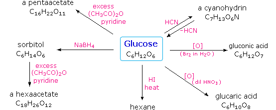
Carbohydrates have been given non-systematic names, although the suffix ose is generally used. The most common carbohydrate is glucose (C6H12O6). Applying the terms defined above, glucose is a monosaccharide, an aldohexose (note that the function and size classifications are combined in one word) and a reducing sugar. The general structure of glucose and many other aldohexoses was established by simple chemical reactions. The following diagram illustrates the kind of evidence considered, although some of the reagents shown here are different from those used by the original scientists.

Hot hydriodic acid (HI) was often used to reductively remove oxygen functional groups from a molecule, and in the case of glucose this treatment gave hexane (in low yield). From this it was concluded that the six carbons are in an unbranched chain. The presence of an aldehyde carbonyl group was deduced from cyanohydrin formation, its reduction to the hexa-alcohol sorbitol, also called glucitol, and mild oxidation to the mono-carboxylic acid, glucuronic acid. Somewhat stronger oxidation by dilute nitric acid gave the diacid, glucaric acid, supporting the proposal of a six-carbon chain. The five oxygens remaining in glucose after the aldehyde was accounted for were thought to be in hydroxyl groups, since a penta-acetate derivative could be made. These hydroxyl groups were assigned, one each, to the last five carbon atoms, because geminal hydroxyl groups are normally unstable relative to the carbonyl compound formed by loss of water. The four middle carbon atoms in the glucose chain are centers of chirality and are colored red.
Glucose and other saccharides are extensively cleaved by periodic acid, thanks to the abundance of vicinal diol moieties in their structure. This oxidative cleavage, known as the Malaprade reaction is particularly useful for the analysis of selective O-substituted derivatives of saccharides, since ether functions do not react. The stoichiometry of aldohexose cleavage is shown in the following equation.

HOCH2(CHOH)4CHO + 5 HIO4   ——> H2C=O + 5 HCO2H + 5 HIO3

The Configuration of Glucose
The four chiral centers in glucose indicate there may be as many as sixteen (24) stereoisomers having this constitution. These would exist as eight diastereomeric pairs of enantiomers, and the initial challenge was to determine which of the eight corresponded to glucose. This challenge was accepted and met in 1891 by the German chemist Emil Fischer. His successful negotiation of the stereochemical maze presented by the aldohexoses was a logical tour de force, and it is fitting that he received the 1902 Nobel Prize for chemistry for this accomplishment. One of the first tasks faced by Fischer was to devise a method of representing the configuration of each chiral center in an unambiguous manner. To this end, he invented a simple technique for drawing chains of chiral centers, that we now call the Fischer projection formula.
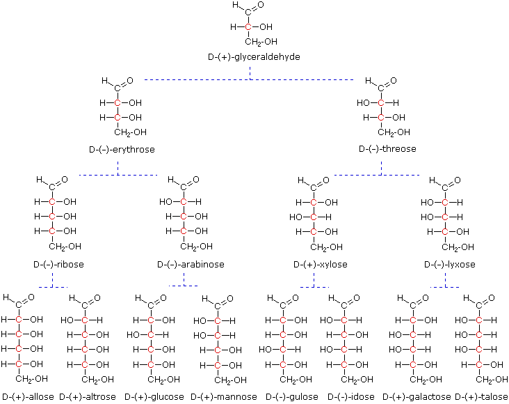
At the time Fischer undertook the glucose project it was not possible to establish the absolute configuration of an enantiomer. Consequently, Fischer made an arbitrary choice for (+)-glucose and established a network of related aldose configurations that he called the D-family. The mirror images of these configurations were then designated the L-family of aldoses. To illustrate using present day knowledge, Fischer projection formulas and names for the D-aldose family (three to six-carbon atoms) are shown below, with the asymmetric carbon atoms (chiral centers) colored red. The last chiral center in an aldose chain (farthest from the aldehyde group) was chosen by Fischer as the D / L designator site. If the hydroxyl group in the projection formula pointed to the right, it was defined as a member of the D-family. A left directed hydroxyl group (the mirror image) then represented the L-family. Fischer’s initial assignment of the D-configuration had a 50:50 chance of being right, but all his subsequent conclusions concerning the relative configurations of various aldoses were soundly based. In 1951 x-ray fluorescence studies of (+)-tartaric acid, carried out in the Netherlands by Johannes Martin Bijvoet (pronounced “buy foot”), proved that Fischer’s choice was correct.
It is important to recognize that the sign of a compound’s specific rotation (an experimental number) does not correlate with its configuration (D or L). It is a simple matter to measure an optical rotation with a polarimeter. Determining an absolute configuration usually requires chemical interconversion with known compounds by stereospecific reaction paths.

Models of representative aldoses  glyceraldehyde, erythrose, threose, ribose, arabinose, allose, altrose, glucose or mannose in the above diagram.

2. Important Reactions

Emil Fischer made use of several key reactions in the course of his carbohydrate studies. These are described here, together with the information that each delivers.

Oxidation
As noted above, sugars may be classified as reducing or non-reducing based on their reactivity with Tollens’, Benedict’s or Fehling’s reagents. If a sugar is oxidized by these reagents it is calledreducing, since the oxidant (Ag(+) or Cu(+2)) is reduced in the reaction, as evidenced by formation of a silver mirror or precipitation of cuprous oxide. The Tollens’ test is commonly used to detect aldehyde functions; and because of the facile interconversion of ketoses and aldoses under the basic conditions of this test, ketoses such as fructose also react and are classified as reducing sugars.

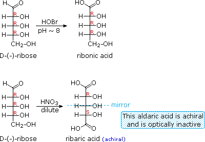
When the aldehyde function of an aldose is oxidized to a carboxylic acid the product is called an aldonic acid. Because of the 2º hydroxyl functions that are also present in these compounds, a mild oxidizing agent such as hypobromite must be used for this conversion (equation 1). If both ends of an aldose chain are oxidized to carboxylic acids the product is called an aldaric acid. By converting an aldose to its corresponding aldaric acid derivative, the ends of the chain become identical (this could also be accomplished by reducing the aldehyde to CH2OH, as noted below). Such an operation will disclose any latent symmetry in the remaining molecule. Thus, ribose, xylose, allose and galactose yield achiral aldaric acids which are, of course, not optically active. The ribose oxidation is shown in equation 2 below.

1.

2.
3.

Other aldose sugars may give identical chiral aldaric acid products, implying a unique configurational relationship. The examples of arabinose and lyxose shown in equation 3 above illustrate this result. Remember, a Fischer projection formula may be rotated by 180º in the plane of projection without changing its configuration.

Reduction
Sodium borohydride reduction of an aldose makes the ends of the resulting alditol chain identical, HOCH2(CHOH)nCH2OH, thereby accomplishing the same configurational change produced by oxidation to an aldaric acid. Thus, allitol and galactitol from reduction of allose and galactose are achiral, and altrose and talose are reduced to the same chiral alditol. A summary of these redox reactions, and derivative nomenclature is given in the following table.

Derivatives of HOCH2(CHOH)nCHO
HOBr Oxidation   ——> HOCH2(CHOH)nCO2H
an Aldonic Acid
HNO3 Oxidation   ——> H2OC(CHOH)nCO2H
an Aldaric Acid
NaBH4 Reduction   ——> HOCH2(CHOH)nCH2OH
an Alditol

Osazone Formation

1.
2.

The osazone reaction was developed and used by Emil Fischer to identify aldose sugars differing in configuration only at the alpha-carbon. The upper equation shows the general form of the osazone reaction, which effects an alpha-carbon oxidation with formation of a bis-phenylhydrazone, known as an osazone. Application of the osazone reaction to D-glucose and D-mannose demonstrates that these compounds differ in configuration only at C-2.

Chain Shortening and Lengthening

1.
2.

These two procedures permit an aldose of a given size to be related to homologous smaller and larger aldoses. The importance of these relationships may be seen in the array of aldose structurespresented earlier, where the structural connections are given by the dashed blue lines. Thus Ruff degradation of the pentose arabinose gives the tetrose erythrose. Working in the opposite direction, a Kiliani-Fischer synthesis applied to arabinose gives a mixture of glucose and mannose. An alternative chain shortening procedure known as the Wohl degradation is essentially the reverse of the Kiliani-Fischer synthesis.

Using these reactions we can now follow Fischer’s train of logic in assigning the configuration of D-glucose.

1.   Ribose and arabinose (two well known pentoses) both gave erythrose on Ruff degradation.
As expected, Kiliani-Fischer synthesis applied to erythrose gave a mixture of ribose and arabinose.
2.   Oxidation of erythrose gave an achiral (optically inactive) aldaric acid. This defines the configuration of erythrose.
3.   Oxidation of ribose gave an achiral (optically inactive) aldaric acid. This defines the configuration of both ribose and arabinose.
4.   Ruff shortening of glucose gave arabinose, and Kiliani-Fischer synthesis applied to arabinose gave a mixture of glucose and mannose.
5.   Glucose and mannose are therefore epimers at C-2, a fact confirmed by the common product from their osazone reactions.
6.   A pair of structures for these epimers can be written, but which is glucose and which is mannose?

In order to determine which of these epimers was glucose, Fischer made use of the inherent C2 symmetry in the four-carbon dissymmetric core of one epimer (B). This is shown in the following diagram by a red dot where the symmetry axis passes through the projection formula. Because of this symmetry, if the aldehyde and 1º-alcohol functions at the ends of the chain are exchanged, epimer B would be unchanged; whereas A would be converted to a different compound.

Fischer looked for and discovered a second aldohexose that represented the end group exchange for the epimer lacking the latent C2 symmetry (A). This compound was L-(+)-gulose, and its exchange relationship to D-(+)-glucose was demonstrated by oxidation to a common aldaric acid product. The remaining epimer is therefore mannose.

3. Ketoses

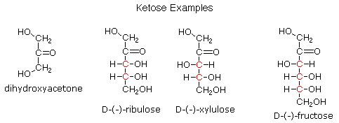
If a monosaccharide has a carbonyl function on one of the inner atoms of the carbon chain it is classified as a ketose. Dihydroxyacetone may not be a sugar, but it is included as the ketose analog of glyceraldehyde. The carbonyl group is commonly found at C-2, as illustrated by the following examples (chiral centers are colored red). As expected, the carbonyl function of a ketose may be reduced by sodium borohydride, usually to a mixture of epimeric products. D-Fructose, the sweetest of the common natural sugars, is for example reduced to a mixture of D-glucitol (sorbitol) and D-mannitol, named after the aldohexoses from which they may also be obtained by analogous reduction. Mannitol is itself a common natural carbohydrate.
Although the ketoses are distinct isomers of the aldose monosaccharides, the chemistry of both classes is linked due to their facile interconversion in the presence of acid or base catalysts. This interconversion, and the corresponding epimerization at sites alpha to the carbonyl functions, occurs by way of an enediol tautomeric intermediate.

Because of base-catalyzed isomerizations of this kind, the Tollens’ reagent is not useful for distinguishing aldoses from ketoses or for specific oxidation of aldoses to the corresponding aldonic acids. Oxidation by HOBr is preferred for the latter conversion.

4. Anomeric Forms of Glucose

Fischer’s brilliant elucidation of the configuration of glucose did not remove all uncertainty concerning its structure. Two different crystalline forms of glucose were reported in 1895. Each of these gave all the characteristic reactions of glucose, and when dissolved in water equilibrated to the same mixture. This equilibration takes place over a period of many minutes, and the change in optical activity that occurs is called mutarotation. These facts are summarized in the diagram below.

When glucose was converted to its pentamethyl ether (reaction with excess CH3I & AgOH), two different isomers were isolated, and neither exhibited the expected aldehyde reactions. Acid-catalyzed hydrolysis of the pentamethyl ether derivatives, however, gave a tetramethyl derivative that was oxidized by Tollen’s reagent and reduced by sodium borohydride, as expected for an aldehyde.

The search for scientific truth often proceeds in stages, and the structural elucidation of glucose serves as a good example. It should be clear from the new evidence presented above, that the open chain pentahydroxyhexanal structure drawn above must be modified. Somehow a new stereogenic center must be created, and the aldehyde must be deactivated in the pentamethyl derivative. A simple solution to this dilemma is achieved by converting the open aldehyde structure for glucose into a cyclic hemiacetal, called a glucopyranose, as shown in the following diagram. The linear aldehyde is tipped on its side, and rotation about the C4-C5 bond brings the C5-hydroxyl function close to the aldehyde carbon. For ease of viewing, the six-membered hemiacetal structure is drawn as a flat hexagon, but it actually assumes a chair conformation. The hemiacetal carbon atom (C-1) becomes a new stereogenic center, commonly referred to as the anomeric carbon, and the α and β-isomers are called anomers.

We can now consider how this modification of the glucose structure accounts for the puzzling facts noted above. First, we know that hemiacetals are in equilibrium with their carbonyl and alcohol components when in solution. Consequently, fresh solutions of either alpha or beta-glucose crystals in water should establish an equilibrium mixture of both anomers, plus the open chain chain form.  Note that despite the very low concentration of the open chain aldehyde in this mixture, typical chemical reactions of aldehydes take place rapidly.
Second, a pentamethyl ether derivative of the pyranose structure converts the hemiacetal function to an acetal. Acetals are stable to base, so this product should not react with Tollen’s reagent or be reduced by sodium borohydride. Acid hydrolysis of acetals regenerates the carbonyl and alcohol components, and in the case of the glucose derivative this will be a tetramethyl ether of the pyranose hemiacetal. This compound will, of course, undergo typical aldehyde reactions.

5. Cyclic Forms of Monosaccharides

As noted above, the preferred structural form of many monosaccharides may be that of a cyclic hemiacetal. Five and six-membered rings are favored over other ring sizes because of their low angle and eclipsing strain. Cyclic structures of this kind are termed furanose (five-membered) or pyranose (six-membered), reflecting the ring size relationship to the common heterocyclic compounds furan and pyran shown on the right. Ribose, an important aldopentose, commonly adopts a furanose structure, as shown in the following illustration. By convention for the D-family, the five-membered furanose ring is drawn in an edgewise projection with the ring oxygen positioned away from the viewer. The anomeric carbon atom (colored red here) is placed on the right. The upper bond to this carbon is defined as beta, the lower bond then is alpha.

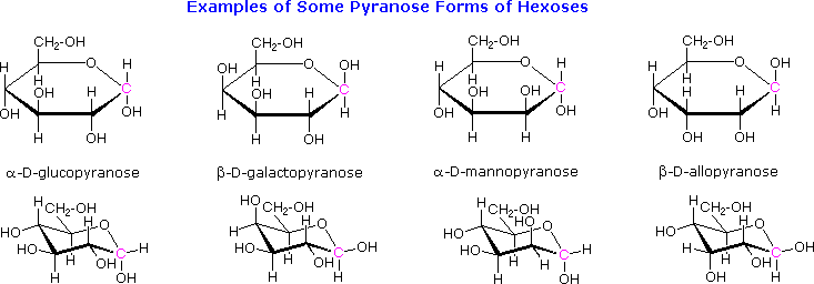
The cyclic pyranose forms of various monosaccharides are often drawn in a flat projection known as a Haworth formula, after the British chemist, Norman Haworth. As with the furanose ring, the anomeric carbon is placed on the right with the ring oxygen to the back of the edgewise view. In the D-family, the alpha and beta bonds have the same orientation defined for the furanose ring (beta is up & alpha is down). These Haworth formulas are convenient for displaying stereochemical relationships, but do not represent the true shape of the molecules. We know that these molecules are actually puckered in a fashion we call a chair conformation. Examples of four typical pyranose structures are shown below, both as Haworth projections and as the more representative chair conformers. The anomeric carbons are colored red.

The size of the cyclic hemiacetal ring adopted by a given sugar is not constant, but may vary with substituents and other structural features. Aldolhexoses usually form pyranose rings and their pentose homologs tend to prefer the furanose form, but there are many counter examples. The formation of acetal derivatives illustrates how subtle changes may alter this selectivity. A pyranose structure for D-glucose is drawn in the rose-shaded box on the left. Acetal derivatives have been prepared by acid-catalyzed reactions with benzaldehyde and acetone. As a rule, benzaldehyde forms six-membered cyclic acetals, whereas acetone prefers to form five-membered acetals. The top equation shows the formation and some reactions of the 4,6-O-benzylidene acetal, a commonly employed protective group. A methyl glycoside derivative of this compound (see below) leaves the C-2 and C-3 hydroxyl groups exposed to reactions such as the periodic acid cleavage, shown as the last step. The formation of an isopropylidene acetal at C-1 and C-2, center structure, leaves the C-3 hydroxyl as the only unprotected function. Selective oxidation to a ketone is then possible. Finally, direct di-O-isopropylidene derivatization of glucose by reaction with excess acetone results in a change to a furanose structure in which the C-3 hydroxyl is again unprotected. However, the same reaction with D-galactose, shown in the blue-shaded box, produces a pyranose product in which the C-6 hydroxyl is unprotected. Both derivatives do not react with Tollens’ reagent. This difference in behavior is attributed to the cis-orientation of the C-3 and C-4 hydroxyl groups in galactose, which permits formation of a less strained five-membered cyclic acetal, compared with the trans-C-3 and C-4 hydroxyl groups in glucose. Derivatizations of this kind permit selective reactions to be conducted at different locations in these highly functional groups. Glycosides

Acetal derivatives formed when a monosaccharide reacts with an alcohol in the presence of an acid catalyst are called glycosides. This reaction is illustrated for glucose and methanol in the diagram below. In naming of glycosides, the “ose” suffix of the sugar name is replaced by “oside”, and the alcohol group name is placed first. As is generally true for most acetals, glycoside formation involves the loss of an equivalent of water. The diether product is stable to base and alkaline oxidants such as Tollen’s reagent. Since acid-catalyzed aldolization is reversible, glycosides may be hydrolyzed back to their alcohol and sugar components by aqueous acid.
The anomeric methyl glucosides are formed in an equilibrium ratio of 66% alpha to 34% beta. From the structures in the previous diagram, we see that pyranose rings prefer chair conformations in which the largest number of substituents are equatorial. In the case of glucose, the substituents on the beta-anomer are all equatorial, whereas the C-1 substituent in the alpha-anomer changes to axial. Since substituents on cyclohexane rings prefer an equatorial location over axial (methoxycyclohexane is 75% equatorial), the preference for alpha-glycopyranoside formation is unexpected, and is referred to as the anomeric effect.

Glycosides abound in biological systems. By attaching a sugar moiety to a lipid or benzenoid structure, the solubility and other properties of the compound may be changed substantially. Because of the important modifying influence of such derivatization, numerous enzyme systems, known as glycosidases, have evolved for the attachment and removal of sugars from alcohols, phenols and amines. Chemists refer to the sugar component of natural glycosides as the glycon and the alcohol component as the aglycon. Salicin, one of the oldest herbal remedies known, was the model for the synthetic analgesic aspirin. A large class of hydroxylated, aromatic oxonium cations called anthocyanins provide the red, purple and blue colors of many flowers, fruits and some vegetables. Peonin is one example of this class of natural pigments, which exhibit a pronounced pH color dependence. The oxonium moiety is only stable in acidic environments, and the color changes or disappears when base is added. The complex changes that occur when wine is fermented and stored are in part associated with glycosides of anthocyanins. Finally, amino derivatives of ribose, such as cytidine play important roles in biological phosphorylating agents, coenzymes and information transport and storage materials.

7. Disaccharides

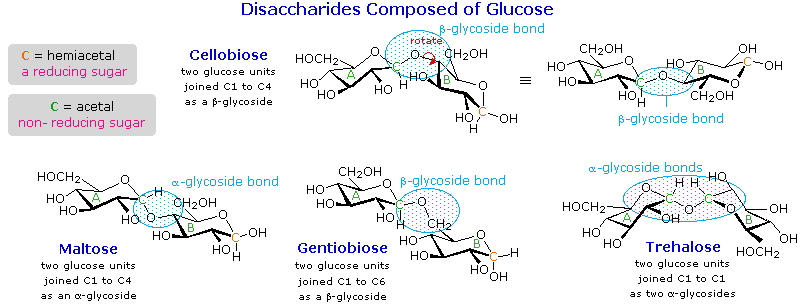
When the alcohol component of a glycoside is provided by a hydroxyl function on another monosaccharide, the compound is called a disaccharide. Four examples of disaccharides composed of two glucose units are shown in the following diagram. The individual glucopyranose rings are labeled A and B, and the glycoside bonding is circled in light blue. Notice that the glycoside bond may be alpha, as in maltose and trehalose, or beta as in cellobiose and gentiobiose. Acid-catalyzed hydrolysis of these disaccharides yields glucose as the only product. Enzyme-catalyzed hydrolysis is selective for a specific glycoside bond, so an alpha-glycosidase cleaves maltose and trehalose to glucose, but does not cleave cellobiose or gentiobiose. A beta-glycosidase has the opposite activity.
In order to draw a representative structure for cellobiose, one of the glucopyranose rings must be rotated by 180º, but this feature is often omitted in favor of retaining the usual perspective for the individual rings. The bonding between the glucopyranose rings in cellobiose and maltose is from the anomeric carbon in ring A to the C-4 hydroxyl group on ring B. This leaves the anomeric carbon in ring B free, so cellobiose and maltose both may assume alpha and beta anomers at that site (the beta form is shown in the diagram). Gentiobiose has a beta-glycoside link, originating at C-1 in ring A and terminating at C-6 in ring B. Its alpha-anomer is drawn in the diagram. Because cellobiose, maltose and gentiobiose are hemiacetals they are all reducing sugars (oxidized by Tollen’s reagent). Trehalose, a disaccharide found in certain mushrooms, is a bis-acetal, and is therefore a non-reducing sugar. A systematic nomenclature for disaccharides exists, but as the following examples illustrate, these are often lengthy.

Cellobiose :   4-O-β-D-Glucopyranosyl-D-glucose (the beta-anomer is drawn)
Maltose :   4-O-α-D-Glucopyranosyl-D-glucose (the beta-anomer is drawn)
Gentiobiose :   6-O-β-D-Glucopyranosyl-D-glucose (the alpha-anomer is drawn)
Trehalose :   α-D-Glucopyranosyl-α-D-glucopyranoside

Although all the disaccharides shown here are made up of two glucopyranose rings, their properties differ in interesting ways. Maltose, sometimes called malt sugar, comes from the hydrolysis of starch. It is about one third as sweet as cane sugar (sucrose), is easily digested by humans, and is fermented by yeast. Cellobiose is obtained by the hydrolysis of cellulose. It has virtually no taste, is indigestible by humans, and is not fermented by yeast. Some bacteria have beta-glucosidase enzymes that hydrolyze the glycosidic bonds in cellobiose and cellulose. The presence of such bacteria in the digestive tracts of cows and termites permits these animals to use cellulose as a food. Finally, it may be noted that trehalose has a distinctly sweet taste, but gentiobiose is bitter.
Disaccharides made up of other sugars are known, but glucose is often one of the components. Lactose, also known as milk sugar, is a galactose-glucose compound joined as a beta-glycoside. It is a reducing sugar because of the hemiacetal function remaining in the glucose moiety. Many adults, particularly those from regions where milk is not a dietary staple, have a metabolic intolerance for lactose. Infants have a digestive enzyme which cleaves the beta-glycoside bond in lactose, but production of this enzyme stops with weaning. Cheese is less subject to the lactose intolerance problem, since most of the lactose is removed with the whey. Sucrose, or cane sugar, is our most commonly used sweetening agent. It is a non-reducing disaccharide composed of glucose and fructose joined at the anomeric carbon of each by glycoside bonds (one alpha and one beta). In the formula shown here the fructose ring has been rotated 180º from its conventional perspective.
8. Polysaccharides

As the name implies, polysaccharides are large high-molecular weight molecules constructed by joining monosaccharide units together by glycosidic bonds. They are sometimes called glycans. The most important compounds in this class, cellulose, starch and glycogen are all polymers of glucose. This is easily demonstrated by acid-catalyzed hydrolysis to the monosaccharide. Since partial hydrolysis of cellulose gives varying amounts of cellobiose, we conclude the glucose units in this macromolecule are joined by beta-glycoside bonds between C-1 and C-4 sites of adjacent sugars. Partial hydrolysis of starch and glycogen produces the disaccharide maltose together with low molecular weight dextrans, polysaccharides in which glucose molecules are joined by alpha-glycoside links between C-1 and C-6, as well as the alpha C-1 to C-4 links found in maltose. Polysaccharides built from other monosaccharides (e.g. mannose, galactose, xylose and arabinose) are also known, but will not be discussed here.

Over half of the total organic carbon in the earth’s biosphere is in cellulose. Cotton fibers are essentially pure cellulose, and the wood of bushes and trees is about 50% cellulose. As a polymer of glucose, cellulose has the formula (C6H10O5)n where n ranges from 500 to 5,000, depending on the source of the polymer. The glucose units in cellulose are linked in a linear fashion, as shown in the drawing below. The beta-glycoside bonds permit these chains to stretch out, and this conformation is stabilized by intramolecular hydrogen bonds. A parallel orientation of adjacent chains is also favored by intermolecular hydrogen bonds. Although an individual hydrogen bond is relatively weak, many such bonds acting together can impart great stability to certain conformations of large molecules. Most animals cannot digest cellulose as a food, and in the diets of humans this part of our vegetable intake functions as roughage and is eliminated largely unchanged. Some animals (the cow and termites, for example) harbor intestinal microorganisms that breakdown cellulose into monosaccharide nutrients by the use of beta-glycosidase enzymes.
Cellulose is commonly accompanied by a lower molecular weight, branched, amorphous polymer called hemicellulose. In contrast to cellulose, hemicellulose is structurally weak and is easily hydrolyzed by dilute acid or base. Also, many enzymes catalyze its hydrolysis. Hemicelluloses are composed of many D-pentose sugars, with xylose being the major component. Mannose and mannuronic acid are often present, as well as galactose and galacturonic acid.

Starch is a polymer of glucose, found in roots, rhizomes, seeds, stems, tubers and corms of plants, as microscopic granules having characteristic shapes and sizes. Most animals, including humans, depend on these plant starches for nourishment. The structure of starch is more complex than that of cellulose. The intact granules are insoluble in cold water, but grinding or swelling them in warm water causes them to burst.
The released starch consists of two fractions. About 20% is a water soluble material called amylose. Molecules of amylose are linear chains of several thousand glucose units joined by alpha C-1 to C-4 glycoside bonds. Amylose solutions are actually dispersions of hydrated helical micelles. The majority of the starch is a much higher molecular weight substance, consisting of nearly a million glucose units, and called amylopectin. Molecules of amylopectin are branched networks built from C-1 to C-4 and C-1 to C-6 glycoside links, and are essentially water insoluble.  The branching in this diagram is exaggerated, since on average, branches only occur every twenty five glucose units.
Hydrolysis of starch, usually by enzymatic reactions, produces a syrupy liquid consisting largely of glucose. When cornstarch is the feedstock, this product is known as corn syrup. It is widely used to soften texture, add volume, prohibit crystallization and enhance the flavor of foods.
Glycogen is the glucose storage polymer used by animals. It has a structure similar to amylopectin, but is even more highly branched (about every tenth glucose unit). The degree of branching in these polysaccharides may be measured by enzymatic or chemical analysis.


Cellulose Nitrate
, first prepared over 150 years ago by treating cellulose with nitric acid, is the earliest synthetic polymer to see general use. The fully nitrated compound, –[C6H7O(ONO2)3]n–, called guncotton, is explosively flammable and is a component of smokeless powder. Partially nitrated cellulose is called pyroxylin. Pyroxylin is soluble in ether and at one time was used for photographic film and lacquers. The high flammability of pyroxylin caused many tragic cinema fires during its period of use. Furthermore, slow hydrolysis of pyroxylin yields nitric acid, a process that contributes to the deterioration of early motion picture films in storage.Cotton, probably the most useful natural fiber, is nearly pure cellulose. The manufacture of textiles from cotton involves physical manipulation of the raw material by carding, combing and spinning selected fibers. For fabrics the best cotton has long fibers, and short fibers or cotton dust are removed. Crude cellulose is also available from wood pulp by dissolving the lignan matrix surrounding it. These less desirable cellulose sources are widely used for making paper.
In order to expand the ways in which cellulose can be put to practical use, chemists have devised techniques for preparing solutions of cellulose derivatives that can be spun into fibers, spread into a film or cast in various solid forms. A key factor in these transformations are the three free hydroxyl groups on each glucose unit in the cellulose chain, –[C6H7O(OH)3]n–. Esterification of these functions leads to polymeric products having very different properties compared with cellulose itself.

Cellulose Acetate, –[C6H7O(OAc)3]n–, is less flammable than pyroxylin, and has replaced it in most applications. It is prepared by reaction of cellulose with acetic anhydride and an acid catalyst. The properties of the product vary with the degree of acetylation. Some chain shortening occurs unavoidably in the preparations. An acetone solution of cellulose acetate may be forced through a spinneret to generate filaments, called acetate rayon, that can be woven into fabrics.
Viscose Rayon, is prepared by formation of an alkali soluble xanthate derivative that can be spun into a fiber that reforms the cellulose polymer by acid quenching. The following general equation illustrates these transformations. The product fiber is called viscose rayon.

ROH NaOH
RO(-) Na(+)   +   S=C=S RO-CS2(-) Na(+) H3O(+)
ROH
cellulose viscose solution rayon

Many complex polysaccharides are found in nature. The galactomannans, consisting of a mannose backbone with galactose side groups, are an interesting and useful example. A (1-4)-linked beta-D-mannose chain is adorned with 1-6-linked alpha-D-galactose units, as shown in the diagram below. The ratio of galactose to mannose usually ranges from 1:2 to 1:4. An important source of this substance is the guar bean, grown principally in northwestern India, and Pakistan. Altough guar protein is not of nutritional value to humans (guar means ‘cow food’ in Hindi), the bean is important as a source of guar gum, a galactomannan which forms a gel in water. The food industry uses this material as a stabilizer in ice cream, cream cheese and salad dressings.


Recently, guar gum has becomes an essential ingredient for mining oil and natural gas in a process called hydraulic fracturing.

CARBOHYDRATES

A carbohydrate is a biological molecule consisting of carbon (C), hydrogen (H) and oxygen (O) atoms, usually with a hydrogen:oxygen atom ratio of 2:1 (as in water); in other words, with the empirical formulaCm(H2O)n (where m could be different from n). Some exceptions exist; for example, deoxyribose, a sugar component of DNA,has the empirical formula C5H10O4. Carbohydrates are technically hydrates of carbon; structurally it is more accurate to view them as polyhydroxy aldehydes and ketones.

The term is most common in biochemistry, where it is a synonym of saccharide, a group that includes sugars, starch, and cellulose. The saccharides are divided into four chemical groups: monosaccharides,disaccharides, oligosaccharides, and polysaccharides. In general, the monosaccharides and disaccharides, which are smaller (lower molecular weight) carbohydrates, are commonly referred to as sugars.[6] The wordsaccharide comes from the Greek word σάκχαρον (sákkharon), meaning “sugar.” While the scientific nomenclature of carbohydrates is complex, the names of the monosaccharides and disaccharides very often end in the suffix -ose. For example, grape sugar is the monosaccharide glucose, cane sugar is the disaccharide sucrose and milk sugar is the disaccharide lactose(see illustration).

Carbohydrates perform numerous roles in living organisms. Polysaccharides serve for the storage of energy (e.g., starch and glycogen) and as structural components (e.g., cellulose in plants and chitin in arthropods). The 5-carbon monosaccharide ribose is an important component of coenzymes (e.g., ATP, FAD andNAD) and the backbone of the genetic molecule known as RNA. The related deoxyribose is a component of DNA. Saccharides and their derivatives include many other important biomolecules that play key roles in the immune system, fertilization, preventing pathogenesis, blood clotting, and development.[7]

In food science and in many informal contexts, the term carbohydrate often means any food that is particularly rich in the complex carbohydrate starch (such ascereals, bread and pasta) or simple carbohydrates, such as sugar (found in candy, jams, and desserts).


Structure

Formerly the name “carbohydrate” was used in chemistry for any compound with the formula Cm (H2O) n. Following this definition, some chemists consideredformaldehyde (CH2O) to be the simplest carbohydrate, while others claimed that title for glycolaldehyde.[9] Today the term is generally understood in the biochemistry sense, which excludes compounds with only one or two carbons.

Natural saccharides are generally built of simple carbohydrates called monosaccharides with general formula (CH2O)n where n is three or more. A typical monosaccharide has the structure H-(CHOH)x(C=O)-(CHOH)y-H, that is, an aldehyde or ketone with many hydroxyl groups added, usually one on each carbonatom that is not part of the aldehyde or ketone functional group. Examples of monosaccharides are glucose, fructose, and glyceraldehydes. However, some biological substances commonly called “monosaccharides” do not conform to this formula (e.g., uronic acids and deoxy-sugars such as fucose) and there are many chemicals that do conform to this formula but are not considered to be monosaccharides (e.g., formaldehyde CH2O and inositol (CH2O)6).

The open-chain form of a monosaccharide often coexists with a closed ring form where the aldehyde/ketone carbonyl group carbon (C=O) and hydroxyl group (-OH) react forming a hemiacetal with a new C-O-C bridge.

Monosaccharides can be linked together into what are called polysaccharides (or oligosaccharides) in a large variety of ways. Many carbohydrates contain one or more modified monosaccharide units that have had one or more groups replaced or removed. For example, deoxyribose, a component of DNA, is a modified version of ribose; chitin is composed of repeating units of N-acetyl glucosamine, a nitrogen-containing form of glucose.

Monosaccharides

Main article: Monosaccharide

D-glucose is an aldohexose with the formula (C·H2O)6. The red atoms highlight thealdehyde group and the blue atoms highlight theasymmetric centerfurthest from the aldehyde; because this -OH is on the right of the Fischer projection, this is a D sugar.

Monosaccharides are the simplest carbohydrates in that they cannot be hydrolyzed to smaller carbohydrates. They are aldehydes or ketones with two or more hydroxyl groups. The general chemical formula of an unmodified monosaccharide is (C•H2O) n, literally a “carbon hydrate.” Monosaccharides are important fuel molecules as well as building blocks for nucleic acids. The smallest monosaccharides, for which n=3, are dihydroxyacetone and D- and L-glyceraldehydes.

Classification of monosaccharides

Alpha-D-glucopyranose-2D-skeletal.pngBeta-D-glucopyranose-2D-skeletal.png

The α and β anomers of glucose. Note the position of the hydroxyl group (red or green) on the anomeric carbon relative to the CH2OH group bound to carbon 5: they either have identical absolute configurations (R,R or S,S) (α), or opposite absolute configurations (R,S or S,R) (β).

Monosaccharides are classified according to three different characteristics: the placement of its carbonylgroup, the number of carbon atoms it contains, and its chiral handedness. If the carbonyl group is analdehyde, the monosaccharide is an aldose; if the carbonyl group is a ketone, the monosaccharide is aketose. Monosaccharides with three carbon atoms are called trioses, those with four are called tetroses, five are called pentoses, six are hexoses, and so on. These two systems of classification are often combined. For example, glucose is an aldohexose (a six-carbon aldehyde), ribose is an aldopentose (a five-carbon aldehyde), and fructose is a ketohexose (a six-carbon ketone).

Each carbon atom bearing a hydroxyl group (-OH), with the exception of the first and last carbons, areasymmetric, making them stereo centers with two possible configurations each (R or S). Because of this asymmetry, a number of isomers may exist for any given monosaccharide formula. Using Le Bel-van’t Hoff rule, the aldohexose D-glucose, for example, has the formula (C·H2O) 6, of which four of its six carbons atoms are stereogenic, making D-glucose one of 24=16 possible stereoisomers. In the case ofglyceraldehydes, an aldotriose, there is one pair of possible stereoisomers, which are enantiomers andepimers. 1, 3-dihydroxyacetone, the ketose corresponding to the aldose glyceraldehydes, is a symmetric molecule with no stereo centers. The assignment of D or L is made according to the orientation of the asymmetric carbon furthest from the carbonyl group: in a standard Fischer projection if the hydroxyl group is on the right the molecule is a D sugar, otherwise it is an L sugar. The “D-” and “L-” prefixes should not be confused with “d-” or “l-“, which indicate the direction that the sugar rotates plane polarized light. This usage of “d-” and “l-” is no longer followed in carbohydrate chemistry.

Ring-straight chain isomerism

Glucose can exist in both a straight-chain and ring form.

The aldehyde or ketone group of a straight-chain monosaccharide will react reversibly with a hydroxyl group on a different carbon atom to form a hemiacetal or hemiketal, forming a heterocyclic ring with an oxygen bridge between two carbon atoms. Rings with five and six atoms are called furanose and pyranose forms, respectively, and exist in equilibrium with the straight-chain form.

During the conversion from straight-chain form to the cyclic form, the carbon atom containing the carbonyl oxygen, called theanomeric carbon, becomes a stereogenic center with two possible configurations: The oxygen atom may take a position either above or below the plane of the ring. The resulting possible pair of stereoisomers is called anomers. In the α anomer, the -OH substituent on the anomeric carbon rests on the opposite side (trans) of the ring from the CH2OH side branch. The alternative form, in which the CH2OH substituent and the anomeric hydroxyl are on the same side (cis) of the plane of the ring, is called theβ anomer.

Use in living organisms

Monosaccharides are the major source of fuel for metabolism, being used both as an energy source (glucose being the most important in nature) and inbiosynthesis. When monosaccharides are not immediately needed by many cells they are often converted to more space-efficient forms, often polysaccharides. In many animals, including humans, this storage form is glycogen, especially in liver and muscle cells. In plants, starch is used for the same purpose. The most abundant carbohydrate, cellulose, is a structural component of the cell wall of plants and many forms of algae. Ribose is a component of RNA. Deoxyribose is a component of DNA. Lyxose is a component of lyxoflavin found in human heart. Ribulose and xylulose occurs in the pentose phosphate pathway. Galactose, a component of milk sugar lactose, is found in galactolipids in plant cell membranes and in glycoproteins in many tissues. Mannose occurs in human metabolism, especially in the glycosylation of certain proteins. Fructose, or fruit sugar, is found in many plants and in humans, it is metabolized in the liver, absorbed directly into the intestines during digestion, and found in semen. Trehalose, a major sugar of insects, is rapidly hydrolyzed into two glucose molecules to support continuous flight.

Disaccharides

Sucrose, also known as table sugar, is a common disaccharide. It is composed of two monosaccharides: D-glucose (left) and D-fructose (right).

Main article: Disaccharide

Two joined monosaccharides are called a disaccharide and these are the simplest polysaccharides. Examples includesucrose and lactose. They are composed of two monosaccharide units bound together by a covalent bond known as aglycosidic linkage formed via a dehydration reaction, resulting in the loss of a hydrogen atom from one monosaccharide and a hydroxyl group from the other. The formula of unmodified disaccharides is C12H22O11. Although there are numerous kinds of disaccharides, a handful of disaccharides are particularly notable.

Sucrose, pictured to the right, is the most abundant disaccharide, and the main form in which carbohydrates are transported in plants. It is composed of one D-glucose molecule and one D-fructose molecule. The systematic name for sucrose, O-α-D-glucopyranosyl-(1→2)-D-fructofuranoside, indicates four things:

  • Its monosaccharides: glucose and fructose
  • Their ring types: glucose is a pyranose and fructose is a furanose
  • How they are linked together: the oxygen on carbon number 1 (C1) of α-D-glucose is linked to the C2 of D-fructose.
  • The -oside suffix indicates that the anomeric carbon of both monosaccharides participates in the glycosidic bond.

Lactose, a disaccharide composed of one D-galactose molecule and one D-glucose molecule, occurs naturally in mammalian milk. The systematic name for lactose is O-β-D-galactopyranosyl-(1→4)-D-glucopyranose. Other notable disaccharides include maltose (two D-glucoses linked α-1,4) and cellulobiose (two D-glucoses linked β-1,4). Disaccharides can be classified into two types.They are reducing and non-reducing disaccharides. If the functional group is present in bonding with another sugar unit, it is called a reducing disaccharide or biose.

Nutrition

Grain products: rich sources of carbohydrates

Carbohydrate consumed in food yields 3.87 calories of energy per gram for simple sugars, and 3.57 to 4.12 calories per gram for complex carbohydrate in most other foods. High levels of carbohydrate are often associated with highly processed foods or refined foods made from plants, including sweets, cookies and candy, table sugar, honey, soft drinks, breads and crackers, jams and fruit products, pastas and breakfast cereals. Lower amounts of carbohydrate are usually associated with unrefined foods, including beans, tubers, rice, and unrefined fruit. Foods from animal carcass have the lowest carbohydrate, but milk does contain lactose.

Carbohydrates are a common source of energy in living organisms; however, no carbohydrate is an essential nutrient in humans. Humans are able to obtain most of their energy requirement from protein and fats, though the potential for some negative health effects of extreme carbohydrate restriction remains, as the issue has not been studied extensively so far. However, in the case of dietary fiber – indigestible carbohydrates which are not a source of energy – inadequate intake can lead to significant increases in mortality.

Following a diet consisting of very low amounts of daily carbohydrate for several days will usually result in higher levels of blood ketone bodies than an isocaloric diet with similar protein content. This relatively high level of ketone bodies is commonly known as ketosis and is very often confused with the potentially fatal condition often seen in type 1 diabetics known as diabetic ketoacidosis. Somebody suffering ketoacidosis will have much higher levels of blood ketone bodies along with high blood sugar, dehydration and electrolyte imbalance.

Long-chain fatty acids cannot cross the blood–brain barrier, but the liver can break these down to produce ketones. However the medium-chain fatty acids octanoic and heptanoic acids can cross the barrier and be used by the brain, which normally relies upon glucose for its energy. Gluconeogenesis allows humans to synthesize some glucose from specific amino acids: from the glycerol backbone in triglycerides and in some cases from fatty acids.

Organisms typically cannot metabolize all types of carbohydrate to yield energy. Glucose is a nearly universal and accessible source of energy. Many organisms also have the ability to metabolize other monosaccharides and disaccharides but glucose is often metabolized first. In Escherichia coli, for example, the lac operonwill express enzymes for the digestion of lactose when it is present, but if both lactose and glucose are present the lac operon is repressed, resulting in the glucose being used first (see: Diauxie). Polysaccharides are also common sources of energy. Many organisms can easily break down starches into glucose, however, most organisms cannot metabolize cellulose or other polysaccharides like chitin and arabinoxylans. These carbohydrate types can be metabolized by some bacteria and protists. Ruminants and termites, for example, use microorganisms to process cellulose. Even though these complex carbohydrates are not very digestible, they represent an important dietary element for humans, called dietary fiber. Fiber enhances digestion, among other benefits.

Based on the effects on risk of heart disease and obesity, the Institute of Medicine recommends that American and Canadian adults get between 45–65% ofdietary energy from carbohydrates. The Food and Agriculture Organization and World Health Organization jointly recommend that national dietary guidelines set a goal of 55–75% of total energy from carbohydrates, but only 10% directly from sugars (their term for simple carbohydrates).

Classification

Nutritionists often refer to carbohydrates as either simple or complex. However, the exact distinction between these groups can be ambiguous. The term complex carbohydrate was first used in the U.S. Senate Select Committee on Nutrition and Human Needs publication Dietary Goals for the United States (1977) where it was intended to distinguish sugars from other carbohydrates (which were perceived to be nutritionally superior). However, the report put “fruit, vegetables and whole-grains” in the complex carbohydrate column, despite the fact that these may contain sugars as well as polysaccharides. This confusion persists as today some nutritionists use the term complex carbohydrate to refer to any sort of digestible saccharide present in a whole food, where fiber, vitamins and minerals are also found (as opposed to processed carbohydrates, which provide energy but few other nutrients). The standard usage, however, is to classify carbohydrates chemically: simple if they are sugars (monosaccharides and disaccharides) and complex if they are polysaccharides (or oligosaccharides).

In any case, the simple vs. complex chemical distinction has little value for determining the nutritional quality of carbohydrates. Some simple carbohydrates (e.g.fructose) raise blood glucose slowly, while some complex carbohydrates (starches), especially if processed, raise blood sugar rapidly. The speed of digestion is determined by a variety of factors including which other nutrients are consumed with the carbohydrate, how the food is prepared, individual differences in metabolism, and the chemistry of the carbohydrate.

The USDA’s Dietary Guidelines for Americans 2010 call for moderate- to high-carbohydrate consumption from a balanced diet that includes six one-ounce servings of grain foods each day, at least half from whole grain sources and the rest from enriched.[32]

The glycemic index (GI) and glycemic load concepts have been developed to characterize food behavior during human digestion. They rank carbohydrate-rich foods based on the rapidity and magnitude of their effect on blood glucose levels. Glycemic index is a measure of how quickly food glucose is absorbed, while glycemic load is a measure of the total absorbable glucose in foods. The insulin index is a similar, more recent classification method that ranks foods based on their effects on blood insulin levels, which are caused by glucose (or starch) and some amino acids in food.

Metabolism

Catabolism

Catabolism is the metabolic reaction which cells undergo to extract energy. There are two major metabolic pathways of monosaccharide catabolism: glycolysis and the citric acid cycle.

In glycolysis, oligo/polysaccharides are cleaved first to smaller monosaccharides by enzymes called glycoside hydrolases. The monosaccharide units can then enter into monosaccharide catabolism. In some cases, as with humans, not all carbohydrate types are usable as the digestive and metabolic enzymes necessary are not present.

Carbohydrate chemistry

Carbohydrate chemistry is a large and economically important branch of organic chemistry. Some of the main organic reactions that involve carbohydrates are: